SDiMatriX
SDi
Matri
X
Equipment
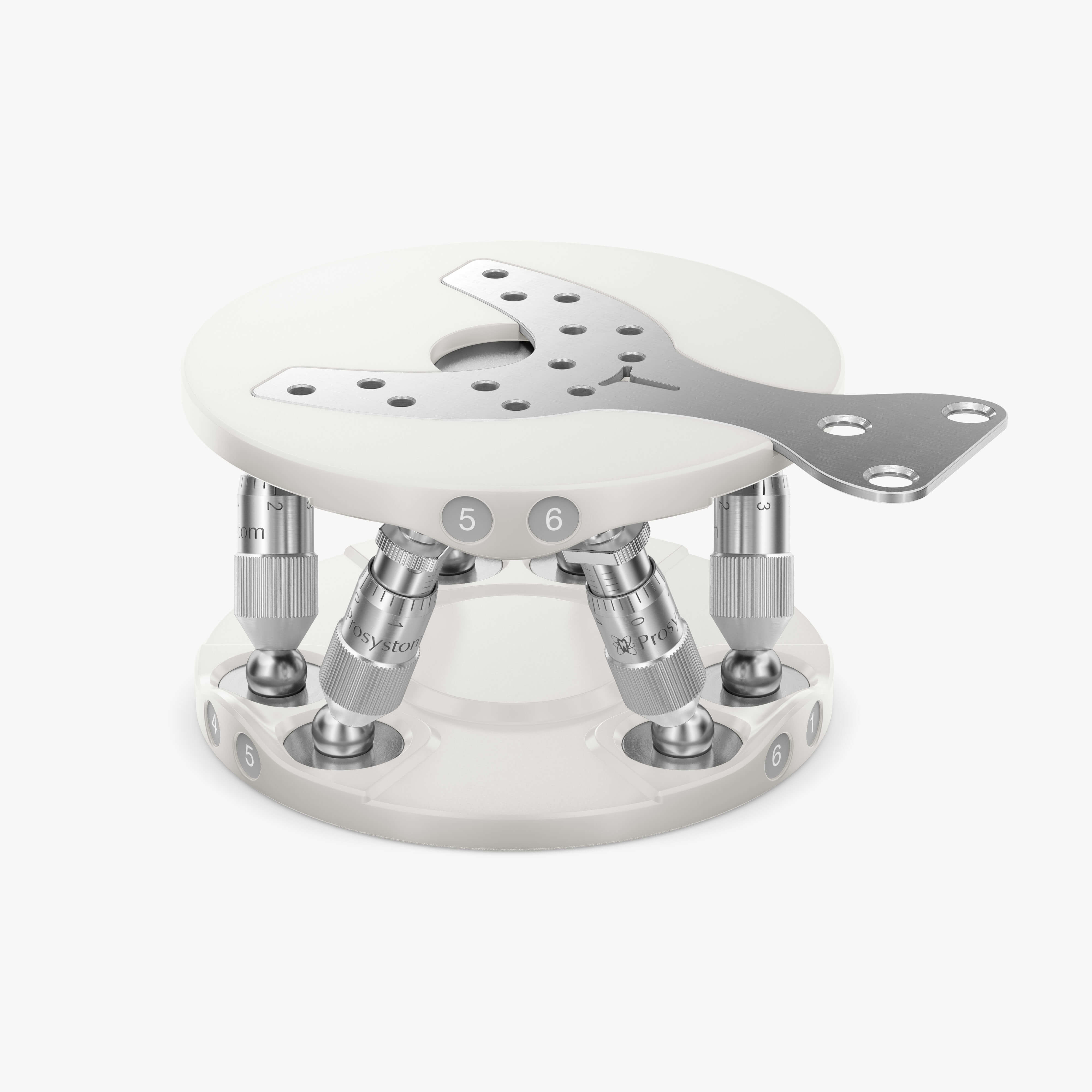
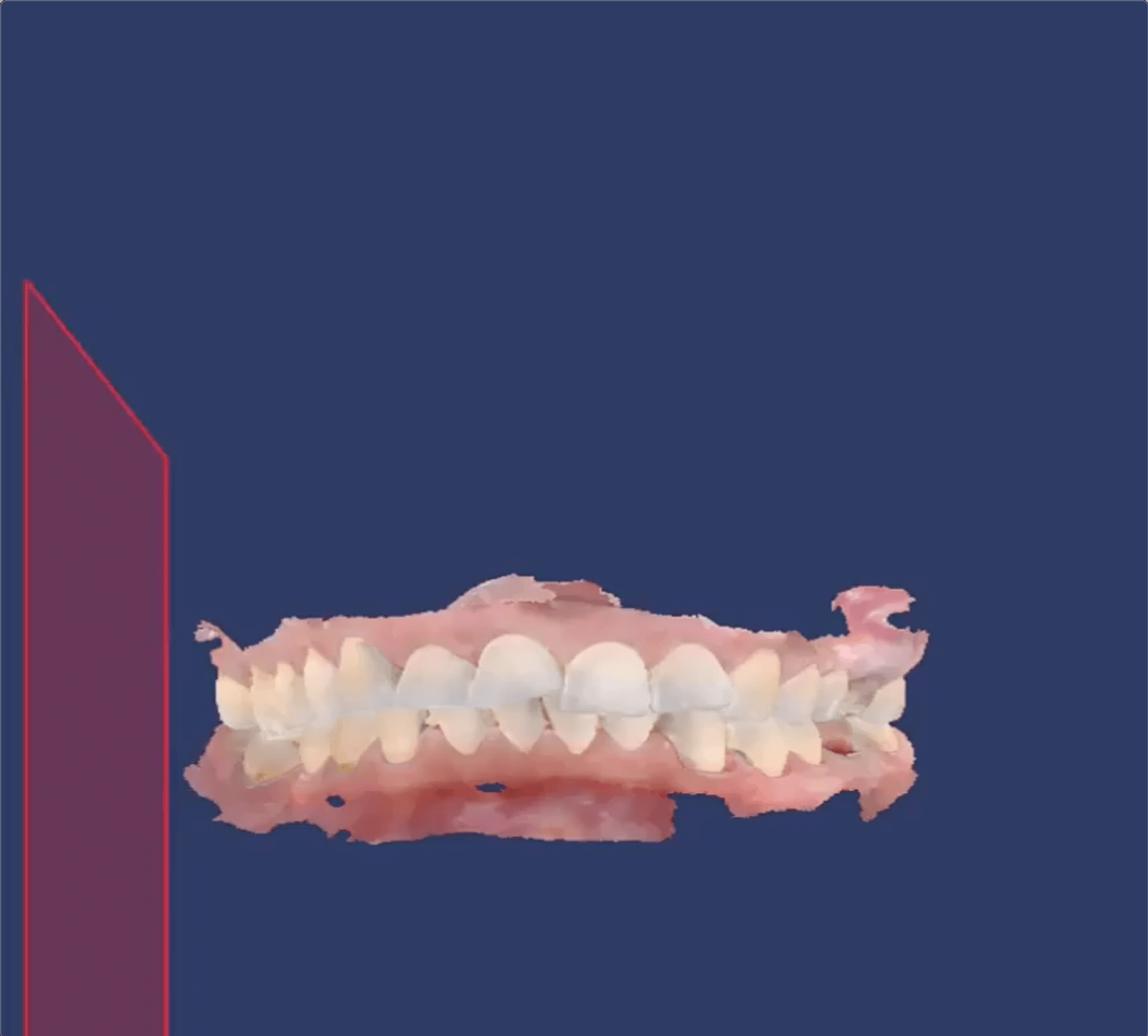
Prostand
3D-stand
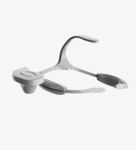
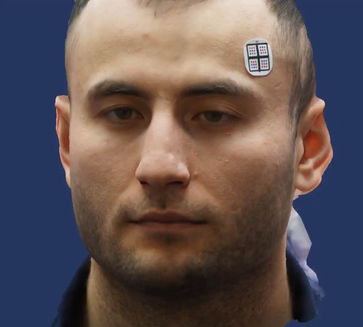
Proarc
Digital facebow
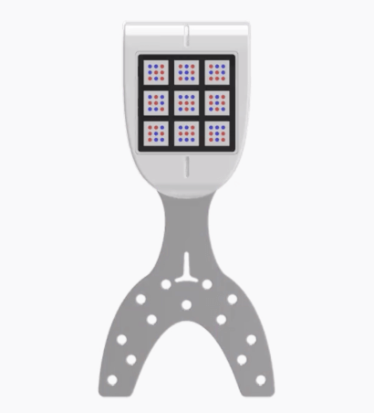
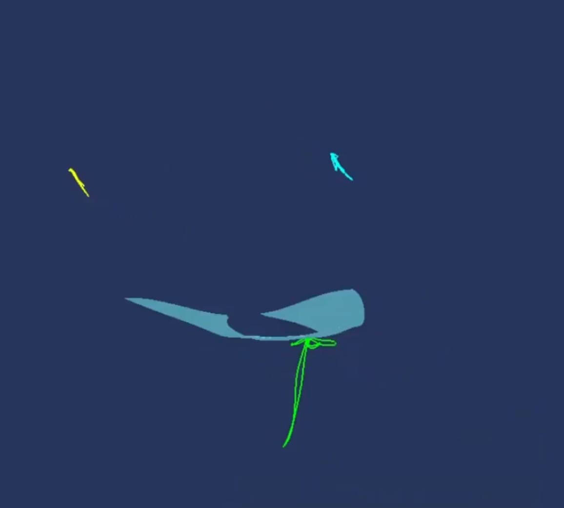
Proaxis
Optical axiograph
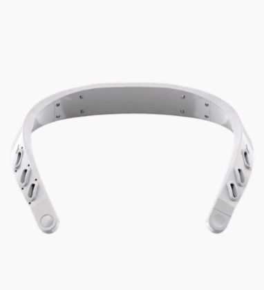
Protens
Wireless Myograph
P-Art
Catalogue
Contacts
slide
1
of 1
slide
1
of 1
Diagnostic center
Equipment
Download PDF catalog
Protens
Wireless myograph
Prostand
3D-stand
Proaxis
Optical Axiograph
Proarc
Proarc
P-Art
Single data space for different diagnostics
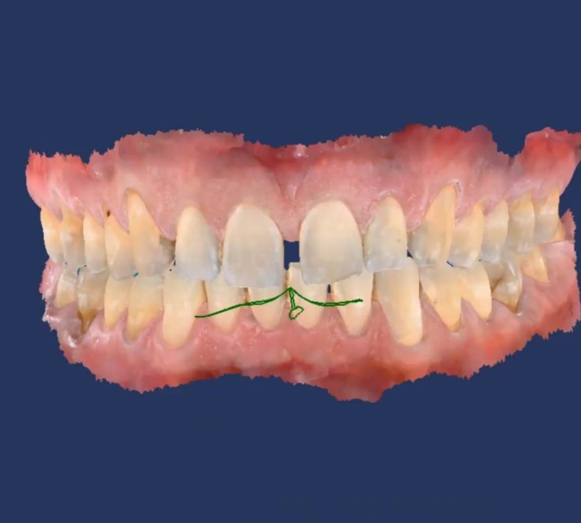
Occlusion analysis
Occlusion analysis
Proline
Teeth alignment with transparent aligners
SDiFace
Facial Analysis
Proshtetics
Orthopedics